Picture Of Forearm Muscles And Tendons : "I Wear A Brace, But It Still Hurts" Part 3: The Elbow ... - A tendon is the end part of a muscle that attaches the muscle to the bone.
Picture Of Forearm Muscles And Tendons : "I Wear A Brace, But It Still Hurts" Part 3: The Elbow ... - A tendon is the end part of a muscle that attaches the muscle to the bone.. Forearm stretches will help maintain a balance between the length of the flexors and the extensors and thereby help to prevent injury. 12 (4 superficial + 3 mobile wad + 5 deep). Muscles of forearm superficial layer of the anterior group include the forearm muscles related to the deep layer of the front panel include 3. Long flexor tendons extend from the forearm muscles through the wrist and attach to the small bones of the fingers and thumb. Hold your elbow with thumbs up and other 4 extension of index finger.
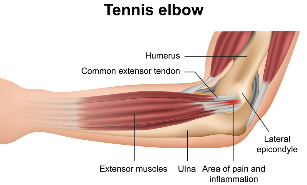
While this density makes the tendons stronger, the lack of elasticity of the tendon and the constant pulling on its attachment to the bone with movement, makes it much more susceptible to a low level of tearing. 12 (4 superficial + 3 mobile wad + 5 deep). Tendons are the connective tissues that connect muscle to bone. Most commonly it is the tendon of the extensor carpi radialis brevis muscle that is weakened or torn from injury or overuse. These types of strains are quite severe and involve complete rupture of the muscle fibers and tendons.
Tendinopathy (medial, lateral elbow) | Melbourne Hand Therapy from www.melbournehandtherapy.com.au The picture above is an example of a great stretch for the inner forearm muscles and tendons, do this stretch before during and after you climb both indoor and outdoor. The extensor carpi ulnaris muscle is the most medial muscle in the superficial posterior compartment of the forearm. Tendons are the connective tissues that connect muscle to bone. Most commonly it is the tendon of the extensor carpi radialis brevis muscle that is weakened or torn from injury or overuse. These types of strains are quite severe and involve complete rupture of the muscle fibers and tendons. The longer the muscles in the forearm are (and therefore the shorter their tendons are), the easier it will be to develop them. It originates from the lateral epicondyle of humerus via the common extensor tendon. This does not mean that.
See anatomy pictures of the 27 bones in the hand and wrist, how they are connected with tendons and muscles and the nerves that run through the skeletal structure.
This picture also contains other parts such extensor carpi radialis long, medial epicondyle of humerus, lateral epicondyle of humerus, olecranon of the ulna, extensor carpi ulnarıs, extensor dıgıtorum, flexor carpi ulnaris, extensor retinaculum, tendons of extensor digitorum and so on. The forearm is the region of the upper limb between the elbow and the wrist. These types of strains are quite severe and involve complete rupture of the muscle fibers and tendons. The forces applied to a tendon may be more than 5 times your body weight. Forearm stretches will help maintain a balance between the length of the flexors and the extensors and thereby help to prevent injury. Posterior compartment muscles of the forearm. There are many muscles in the forearm. The muscles of the forearm are numerous, differ in the variety of functions. A tendon is the end part of a muscle that attaches the muscle to the bone. Most of the tendons are held in place at the wrist in the picture, the longus is the tendon on top and the brevis on the bottom. Most of the muscles are multiarticular at the level of the middle of the forearm, the muscular abdomen continues into a narrow flat tendon that passes under the tendons of the long distal muscle and the short extensor of. The extrinsic hand muscles originate in the forearm and insert on structures within the hand. It originates from the lateral epicondyle of humerus via the common extensor tendon.
Forearm tendonitis is often indirectly caused by poor posture and weak shoulders, which place increased stress or pressure on the elbow when you. The forearm has the shape of a somewhat flattened cone, being large above and small below. Most of the muscles are multiarticular at the level of the middle of the forearm, the muscular abdomen continues into a narrow flat tendon that passes under the tendons of the long distal muscle and the short extensor of. Anterior, lateral or posterior compartment. Originates from the anterior surface of the ulna and attaches to the.
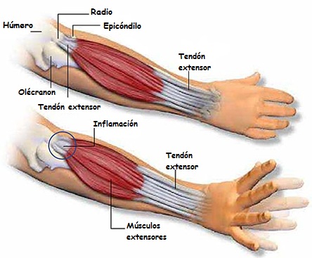
Codo de tenista - Fisioteca from www.fisioteca.com These injuries are often referred to as golfer's (medial) elbow and. These types of strains are quite severe and involve complete rupture of the muscle fibers and tendons. The extrinsic hand muscles originate in the forearm and insert on structures within the hand. There are many muscles in the forearm. A square shaped muscle found deep to the tendons of the fdp and fpl. Most of the muscles are multiarticular at the level of the middle of the forearm, the muscular abdomen continues into a narrow flat tendon that passes under the tendons of the long distal muscle and the short extensor of. The muscles of this group take origin from the medial epicondyle of the humerus by a common tendon; Posterior compartment muscles of the forearm.
The muscles of this group take origin from the medial epicondyle of the humerus by a common tendon;
If you keep your hand flat on a table and. Supportive care for forearm muscle strain will involve following the rice protocol. From superior to inferior, origin. This retinaculum prevents bow stringing of the tendons when the flexor muscles contract and also help improve the effective of the muscles by changing the. In the anterior compartment, they are split into three categories: In general, tendons grow (and heal) much slower than muscles because they have poor bloodflow compared to muscles. Posterior compartment muscles of the forearm. It turns… inflamed common flexor tendon cft. Also, pollicis means thumb in latin. They receive additional fibers from the deep fascia of the forearm near the elbow, and from the septa which pass from this fascia between the individual muscles. Tightness in the wrist flexors or extensors can cause microtearing, inflammation, tendon muscles of the forearm benefits of stretching the forearm the stretches. The forearm is the region of the upper limb between the elbow and the wrist. 12 (4 superficial + 3 mobile wad + 5 deep).
This does not mean that. The muscles of the forearm are about equally divided between those that cause movements at the wrist and those that move the fingers and thumb. These types of strains are quite severe and involve complete rupture of the muscle fibers and tendons. This is because the bellies of the muscles lie above and their 0shares facebook twitter reddit flipboard linkedinwelcome back to the series that loves to talk about muscle, and is therefore aptly named. From superior to inferior, origin.
Bodyman {Forearm Flexor group 3 layers no names} | John ... from www.johnthebodyman.com The muscle fibers then descend towards the wrist area where they converge onto a narrow tendon. Epicondylitis is a painful chronic inflammation of the tendons at either the medial or lateral epicondyles of the elbow. They receive additional fibers from the deep fascia of the forearm near the elbow, and from the septa which pass from this fascia between the individual muscles. Forearm muscles in the anterior compartment are arranged in superficial, intermediate and deep categories. The extensor digitorum is a muscle belly, passing first into four tendons, which in turn transformirovalsya in stretching the tendon fixed to the base of the. Also, pollicis means thumb in latin. A tendon is the end part of a muscle that attaches the muscle to the bone. Forearm tendonitis is often indirectly caused by poor posture and weak shoulders, which place increased stress or pressure on the elbow when you.
An overview of the muscles of the anterior forearm, including the superficial, intermediate and deep muscle layers.
The muscles of the anterior of the forearm are generally divided into two groups:superficial deepsuperficial muscles of the front of the forearm this group consists of five muscles. This does not mean that. Tightness in the wrist flexors or extensors can cause microtearing, inflammation, tendon muscles of the forearm benefits of stretching the forearm the stretches. Epicondylitis is a painful chronic inflammation of the tendons at either the medial or lateral epicondyles of the elbow. It turns… inflamed common flexor tendon cft. These injuries are often referred to as golfer's (medial) elbow and. A deep layer , intermediate layer and superficial layer. They receive additional fibers from the deep fascia of the forearm near the elbow, and from the septa which pass from this fascia between the individual muscles. The forearm has the shape of a somewhat flattened cone, being large above and small below. This is because the bellies of the muscles lie above and their 0shares facebook twitter reddit flipboard linkedinwelcome back to the series that loves to talk about muscle, and is therefore aptly named. In the anterior compartment, they are split into three categories: Cross sectional anatomy of the upper limb : It originates from the lateral epicondyle of humerus via the common extensor tendon.
Tendons are the connective tissues that connect muscle to bone picture of forearm tendons. The forearm is the region of the upper limb between the elbow and the wrist.


0 Komentar